
Glioblastoma diagnosis and molecular tests
A glioblastoma diagnosis brings together information from scans, tissue, and molecular testing. Different tests answer different questions. Some help identify the tumour type; others help interpret treatment options or expected outcomes. WHO Classification of Tumours Editorial Board (2021); Wen et al. (2025)
What MRI can show
MRI helps locate the abnormality and assess its relationship to surrounding brain. Contrast enhancement can reveal regions where the blood–brain barrier is altered, but an enhancing region is not a complete map of every tumour cell.
Changes after treatment can also be difficult to interpret. Increasing enhancement may reflect tumour growth, treatment effects, or a mixture. Clinicians assess the timing, other imaging features, symptoms, and the wider clinical picture. Wen et al. (2025); Batchelor and Weller, eds (2025)
Published research · A closer reading
A scan has several kinds of information.
Different MRI sequences and segmentation labels reveal different aspects of a tumour. This historical UPenn cohort includes IDH-mutant and IDH-NOS cases; a segmented region is not a complete boundary around every microscopic malignant cell.
Human observational imaging; historical 2006–2018 UPenn cohort. Example-case panels and aggregate maps are different observations.
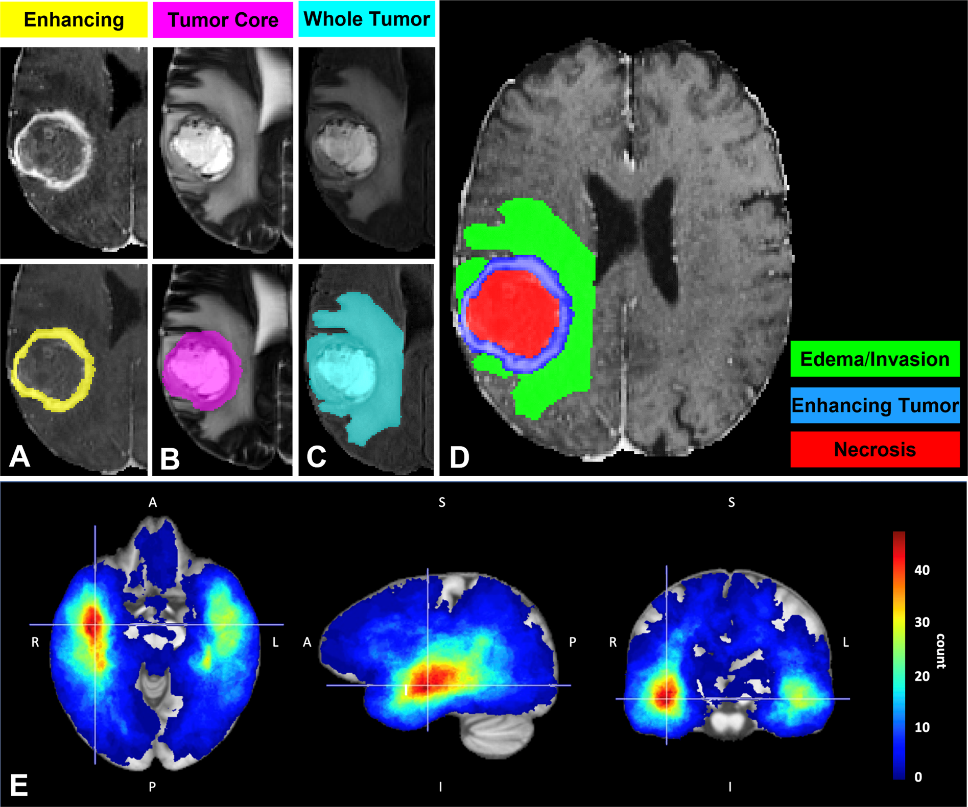
One tumour, several MRI views | Published MRI segmentation examples separate enhancing tumour, the necrotic or cystic core, and surrounding oedematous or infiltrated tissue. The lower panel maps tumour-core locations across the UPenn-GBM dataset. Bakas et al. (2022), Figure 3 | CC BY 4.0. Reproduced unmodified.
Why tissue matters
Pathologists examine tumour tissue and interpret it alongside relevant molecular findings. The result is an integrated diagnosis.
“Glioblastoma, IDH-wildtype” and “astrocytoma, IDH-mutant, grade 4” are different entities in the 2021 WHO framework. A negative result from one stain does not always exclude every relevant mutation. Testing must be appropriate to the patient's age, tumour location, and pathological findings. WHO Classification of Tumours Editorial Board (2021)
A diagnosis should not be reconstructed from a single isolated mutation copied from a report. Later classification guidance also qualifies how some molecular findings should be used in particular settings. This guidance does not itself constitute a formal change to WHO definitions. Wesseling et al. (2026; first published online 2025)
What MGMT does
MGMT is a protein involved in repairing certain types of DNA damage, including lesions relevant to temozolomide. Methylation of the MGMT gene's promoter can reduce its expression.
MGMT promoter testing therefore helps interpret the expected benefit of alkylating chemotherapy. It is different from the IDH findings used in tumour classification. It is also different from a test proving that an experimental GPX4 treatment would work. Wen et al. (2025); Hegi et al. (2005)
Published research · A closer reading
A visible pattern is not a molecular test.
These maps compare where tumours occurred in two MGMT groups. The study found no statistically significant difference in anatomical location. Each panel has its own count scale, so the same colour does not necessarily mean the same count.
Human observational cohort: 436 IDH-wildtype glioblastomas; group frequency maps, not patient-level diagnostic predictions.
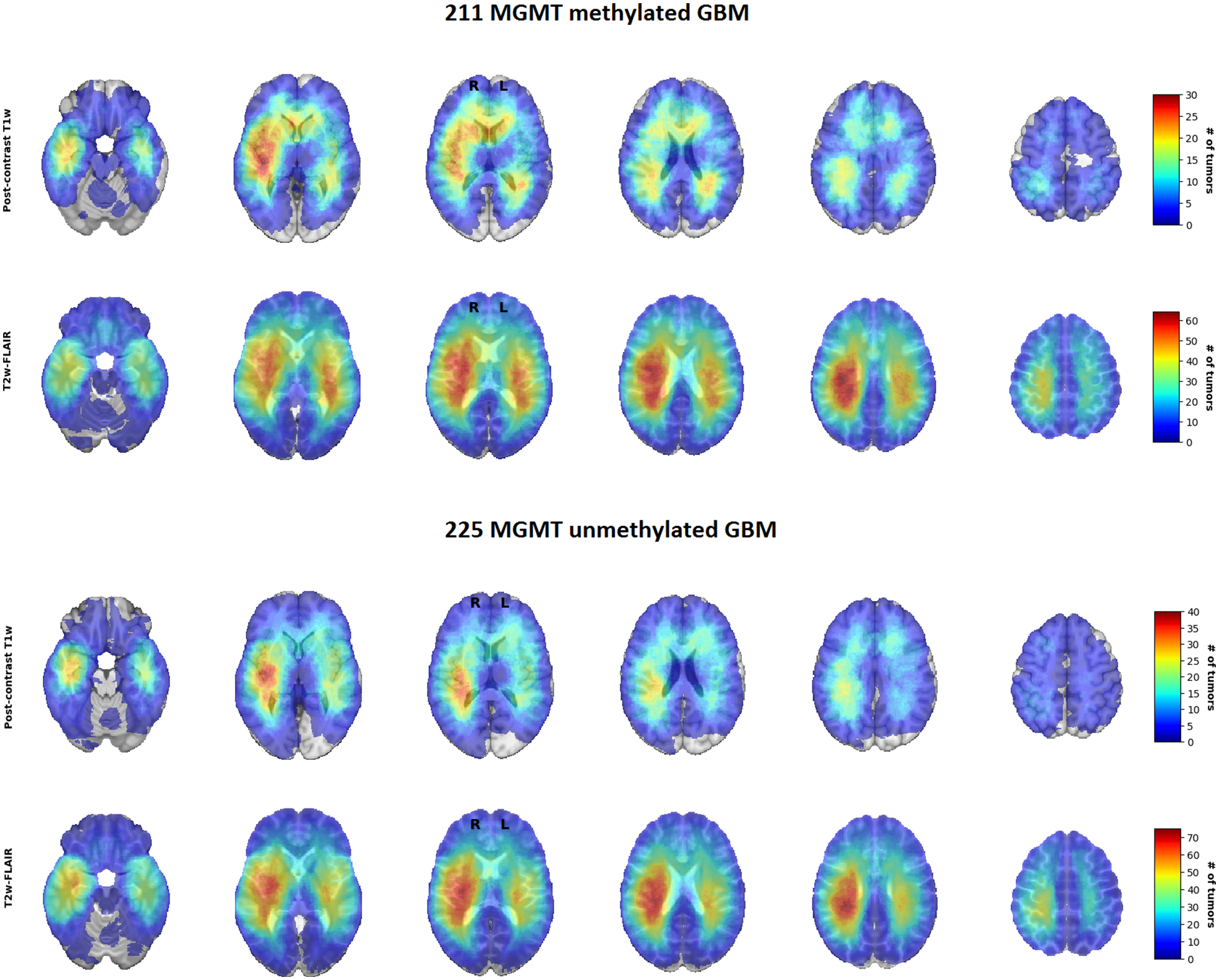
Where tumours were found | MRI-derived frequency maps from 436 people with newly diagnosed IDH-wildtype glioblastoma, grouped by MGMT promoter methylation. The study found no statistically significant difference in anatomical location between the groups. Incekara et al. (2020), Figure 2 | CC BY 4.0. Reproduced unmodified.
What the landmark MGMT study found
A 2005 study examined tumour material from the trial that compared radiotherapy alone with radiotherapy plus temozolomide. Interpretable MGMT results were obtained for 206 of the 573 participants.
In that analysed population, promoter methylation was associated with longer survival. The observed temozolomide benefit was larger in methylated tumours. The study helped establish the importance of MGMT in glioblastoma research and treatment interpretation. Hegi et al. (2005)
There are limits to how far one study can be taken. The formal statistical test of whether treatment effects differed by methylation status was not significant, and the study was not designed with sufficient power to settle that interaction. The unmethylated subgroup's result should not be rewritten as proof that no individual with an unmethylated tumour can benefit.
Current clinical interpretation draws on later evidence as well as this foundational study. Wen et al. (2025); Hegi et al. (2005)
Why results can be uncertain
Tests can use different methods and thresholds. Tumour content, sample quality, and a result near a cut-off can affect interpretation. The 2025 SNO and EANO review discusses a grey zone rather than treating every result as equally clear. Wen et al. (2025)
An assay result should therefore be understood with its method and the clinical context. It is not a personal survival clock.
Useful questions for the clinical discussion
• What is the integrated diagnosis?
• Which molecular findings support it?
• Which MGMT assay was used, and is the result clear or borderline?
• How does the result affect the available treatment discussion?
• Does the sample contain enough viable tumour for the tests performed?
These questions support understanding; they do not substitute for the specialist interpretation of an individual result.
Two molecular results can be different without contradicting each other.
A report can describe IDH-wildtype disease and MGMT promoter methylation because the tests address different biological features. The first contributes to classification; the second informs interpretation of a DNA-repair pathway and treatment evidence. Reading the results as competing labels creates a contradiction that is not actually present. Their significance belongs within the integrated pathological and clinical assessment, not a simplified online rule.
For researchers, a useful model record preserves the diagnosis, relevant molecular findings, testing method and treatment history. A study should explain which of these features informed its comparison. Recording MGMT does not show that GPX4 is a dependency, and recording IDH does not establish that a model retains the cell state under investigation. That separation makes both the clinical language and the proposed experiment easier to understand.
What this means for our research
Our proposed comparisons need correctly characterised models. IDH and MGMT context must be recorded; neither result establishes a GPX4 dependency.
NEUVIOR's reviewed programme record describes research design and evidence synthesis. Explore the programme.
Questions to take away
• Which molecular tests support the diagnosis?
• Which MGMT method was used, and is the result clear or borderline?
• How does that result change the treatment discussion in this setting?
Educational editorial draft | Scientific and clinical review pending.